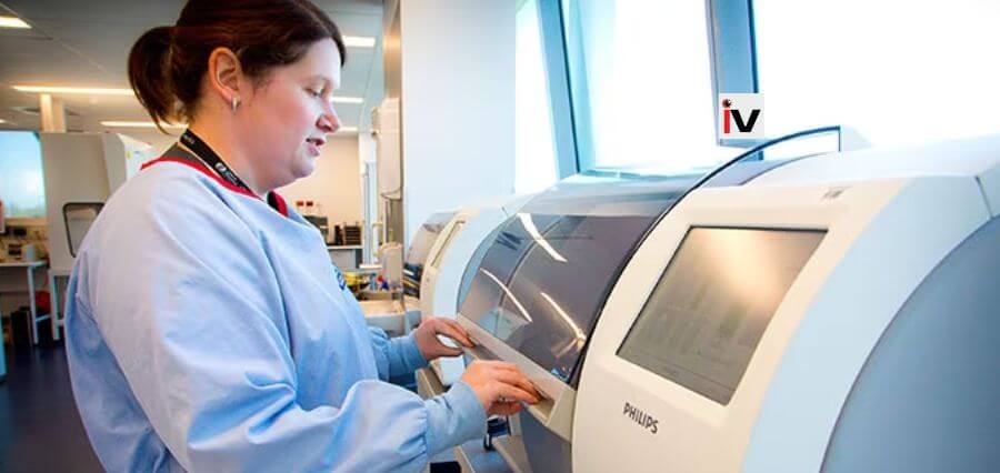
After a ten-year research initiative that is expected to revolutionise medical diagnosis, patients will receive quicker and more accurate pathology reports.
A microscope scanning and analysis system that has been automated in Brisbane by the University of Queensland and Sullivan Nicolaides Pathology (SNP) has been tested, put into use, and accredited in advance of widespread implementation.
Brian Lovell, a professor of AI at UQ, said the approach greatly increased test costs, quality, and speed.
The National Association of Testing Authorities (NATA) has validated this digital pathology system, which conducts hundreds of tests each day, according to Professor Lovell.
“The system can sometimes boost pathologists’ and scientists’ productivity by a factor of ten or more.”
The method also makes it possible to use telepathology to get second opinions. It also vastly improves record keeping and access to historical records because glass slides are no longer need to be stored for years.
The technology, according to SNP Chief Executive Officer Dr. Michael Harrison, is a game changer in many facets of healthcare.
The technique is already being used by SNP laboratories in Brisbane to increase diagnosis accuracy and speed, according to Dr. Harrison. Our scientists no longer spend hours shackled to a microscope; instead, they use digital images, frequently with related AI.
This represents the biggest shift in morphological test performance in decades.
According to Professor Lovell, capturing clear, in-focus photographs without the assistance of a human has historically presented significant challenges.
“Digital pathology images are often thousands of times larger than typical digital photos,” he said.
Adding further he said, “This had meant microscopy for diagnosing from tissue, blood and other specimen types was unable to be automated until now.”
Read More: Click here

